NAIAEANIBL
NAIAEANIBL
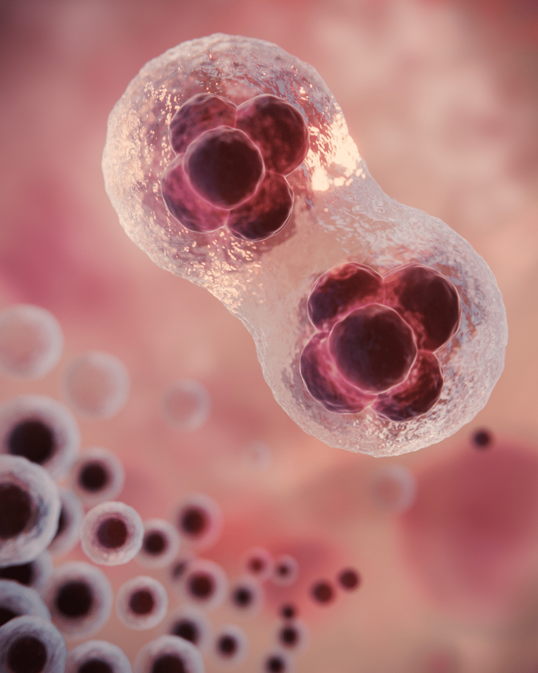
New Artificial Intelligence Algorithm for Non-Invasive Automatic Evaluation of Blastocyst
Improving embryo evaluation is essential to increasing the success of assisted reproduction treatments, and NAIAEANIBL focuses on doing so through safe, non-invasive approaches that reduce risks and improve outcomes for patients.

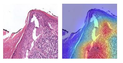
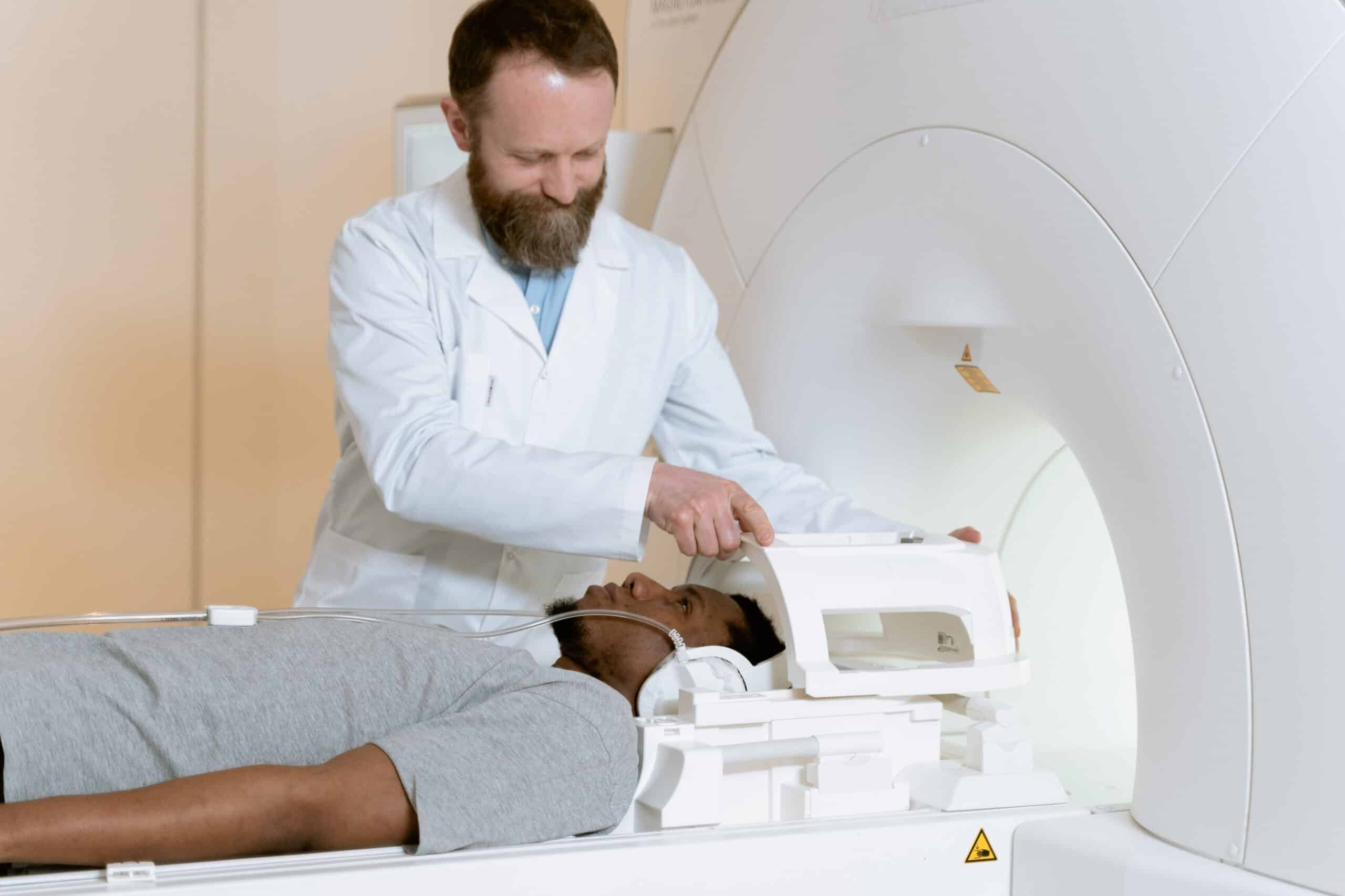
Embryo evaluation is one of the most critical aspects of assisted reproduction treatments, as it largely determines the success of the process. Currently, techniques such as Preimplantation Genetic Testing make it possible to analyze the chromosomal status of embryos, but they require invasive, costly procedures with important limitations. In this context, the NAIAEANIBL project aims to move towards a new generation of diagnostic tools that are more precise, accessible, and respectful of the embryo.
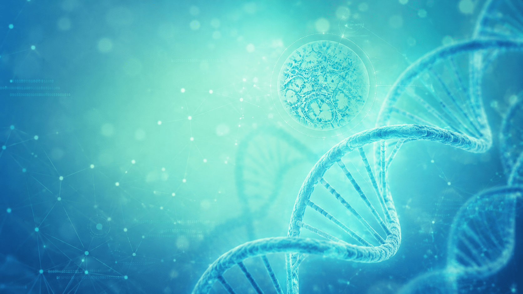
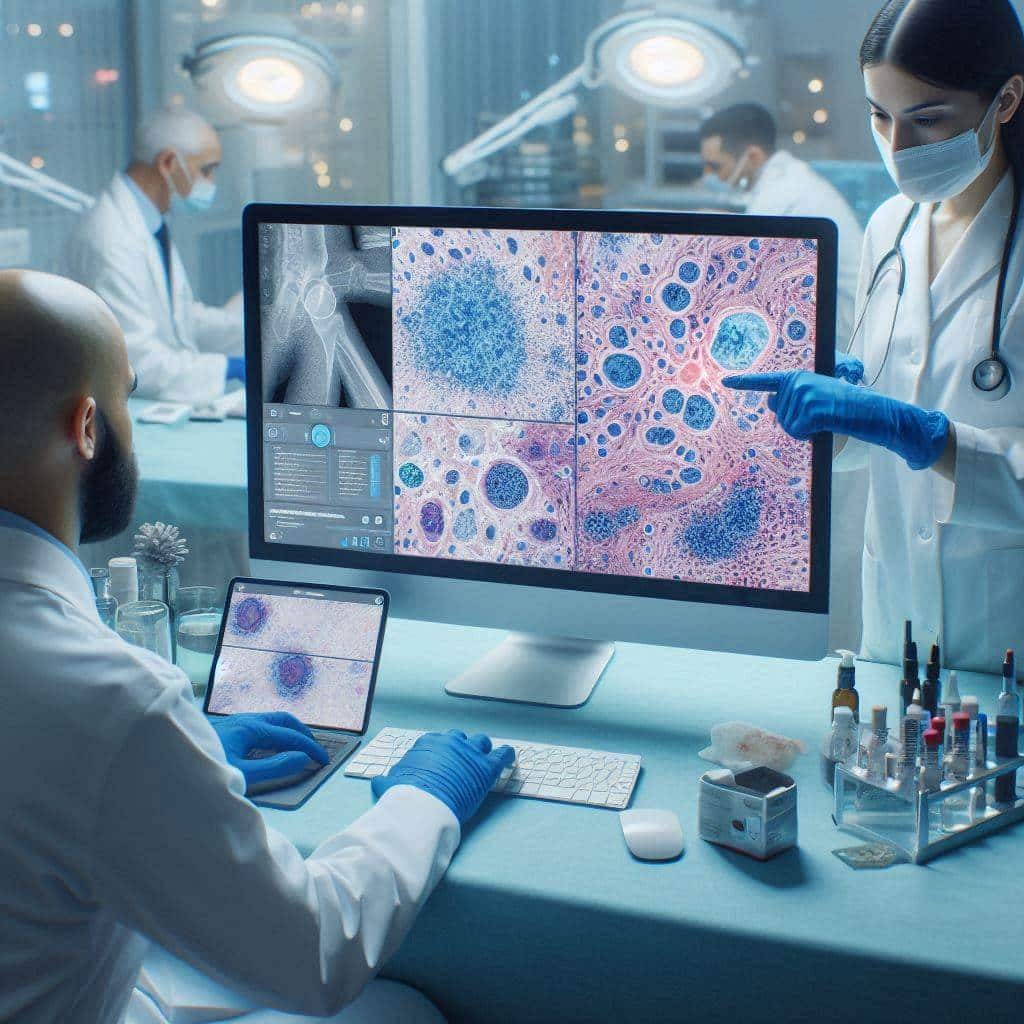
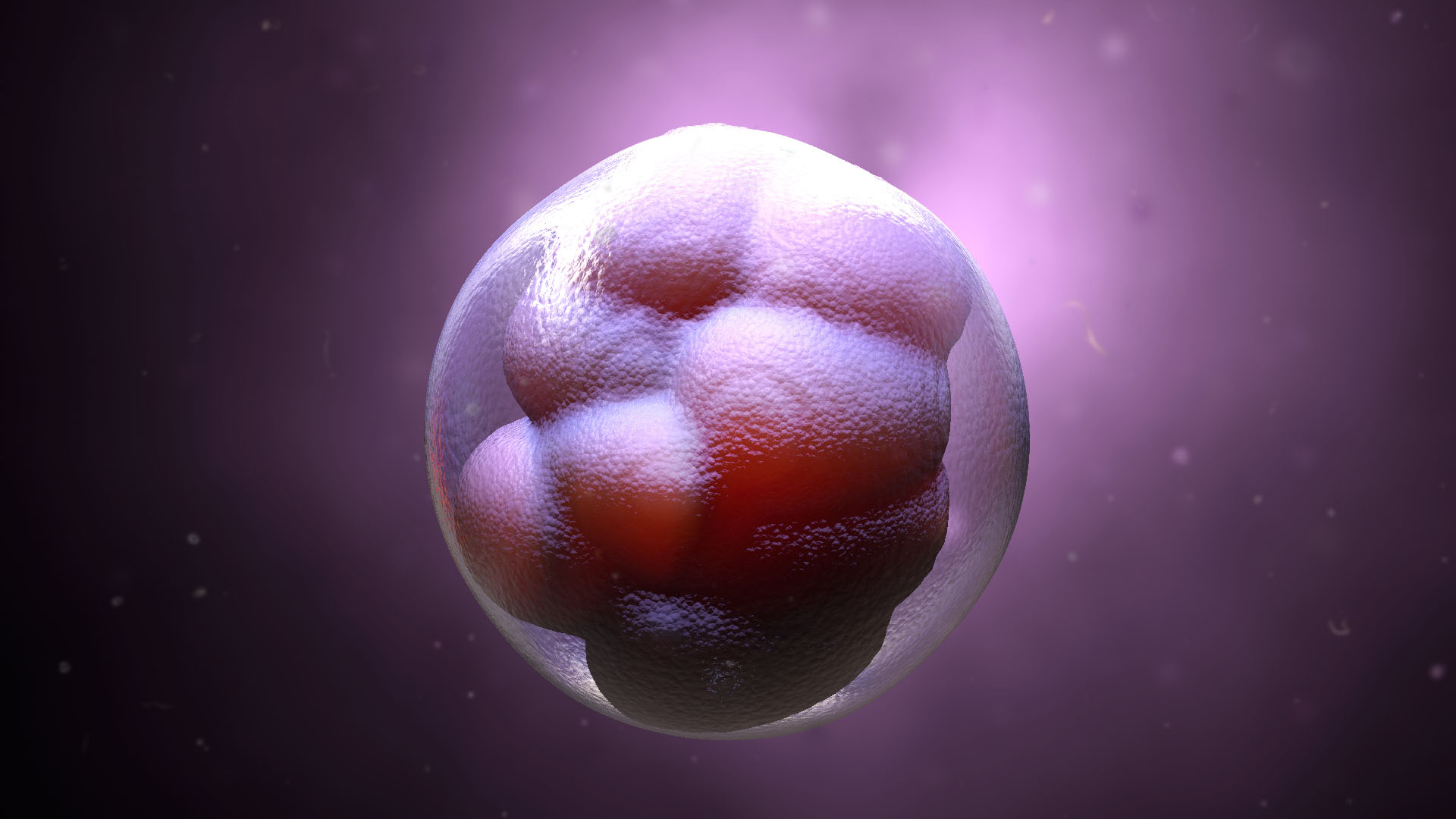
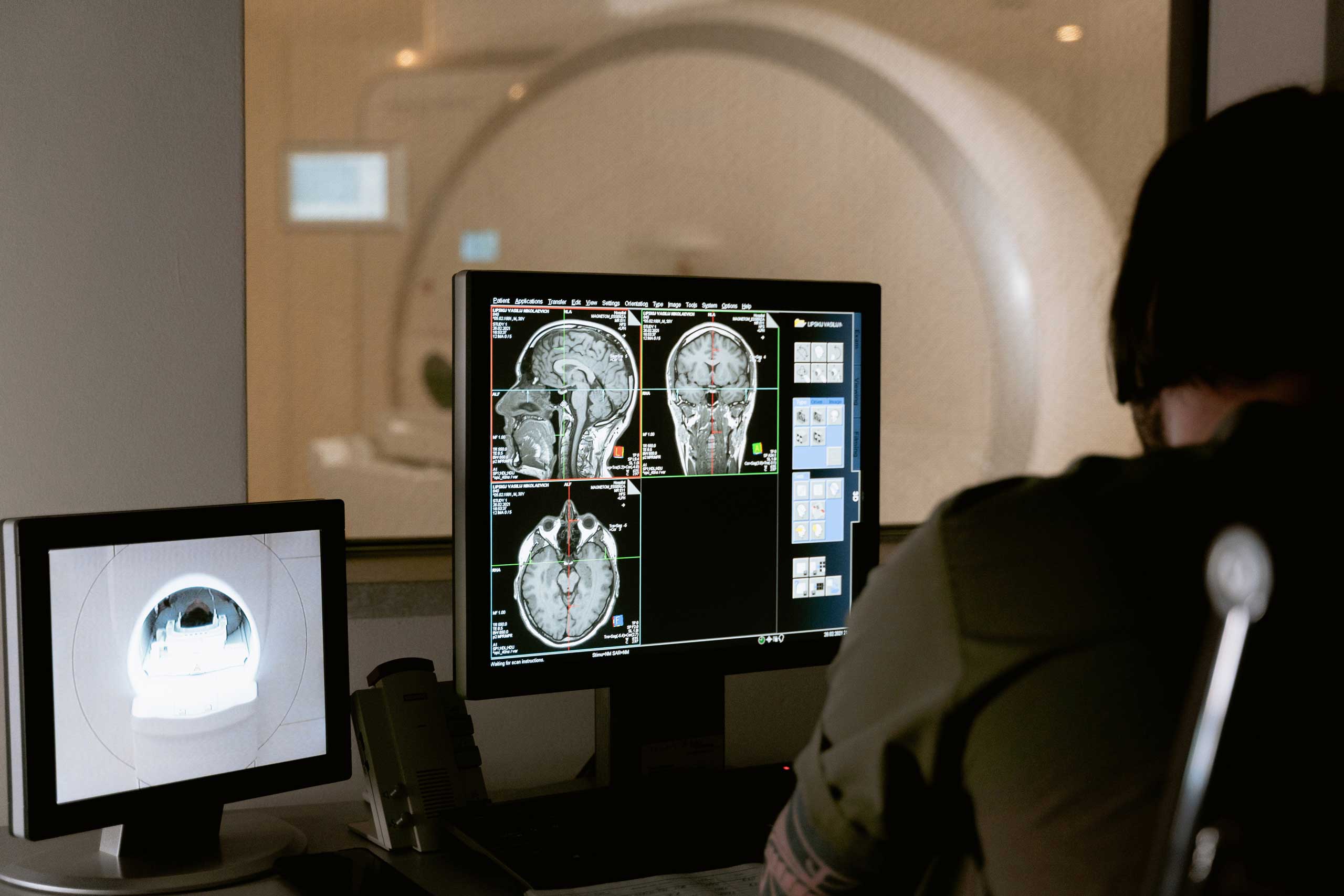
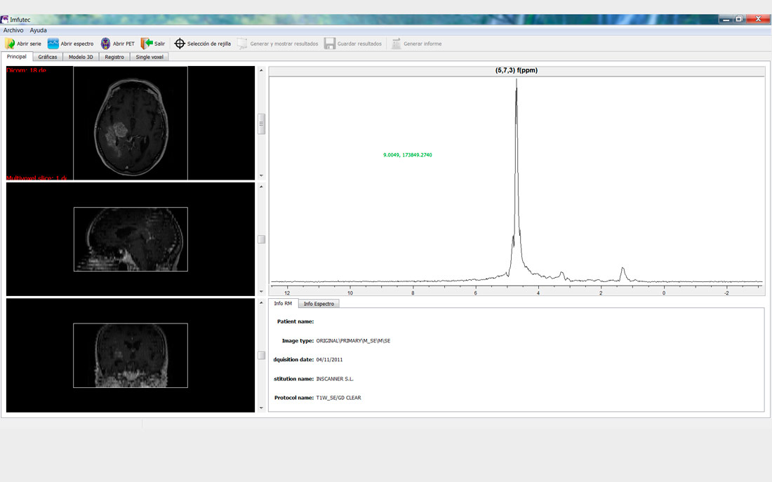
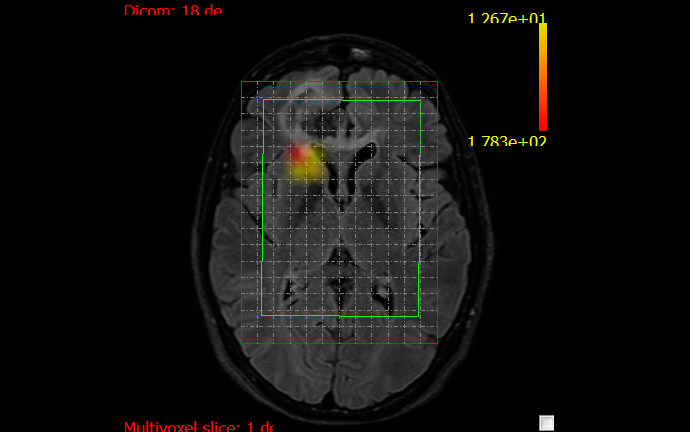
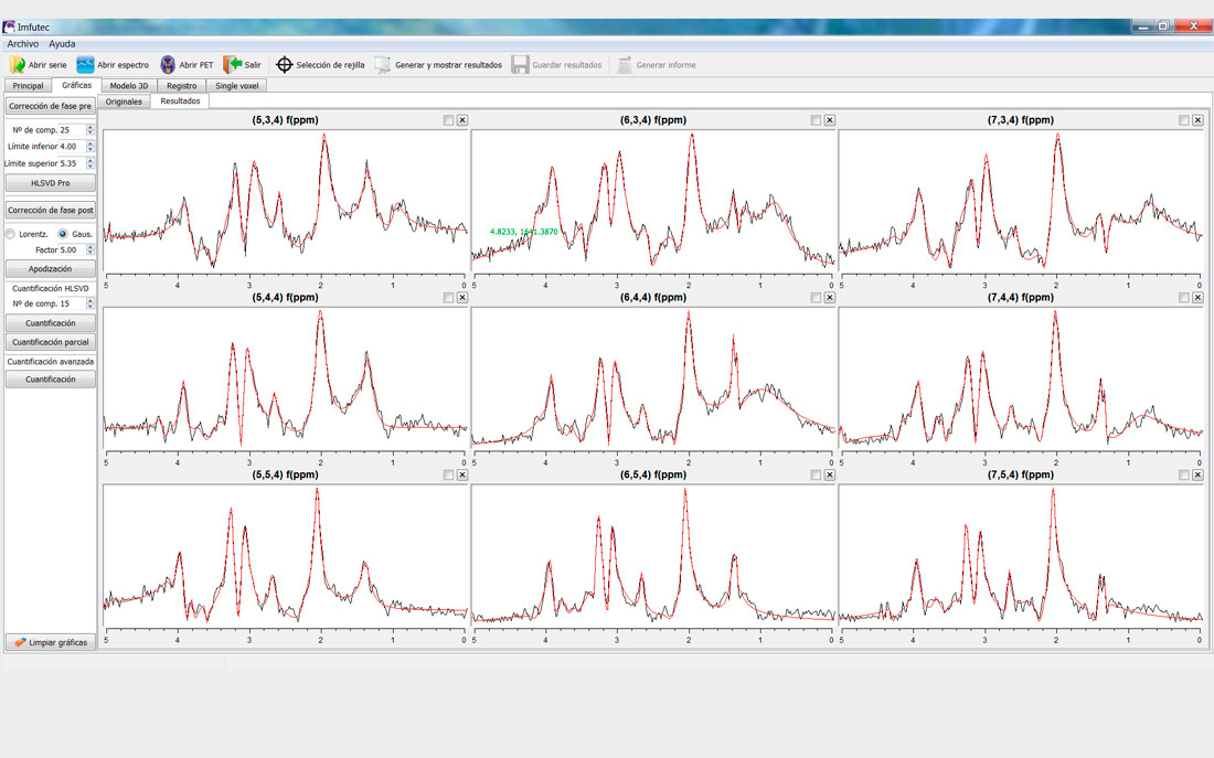
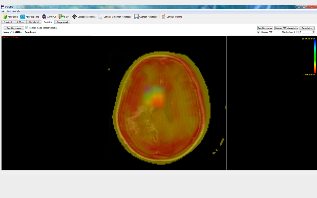
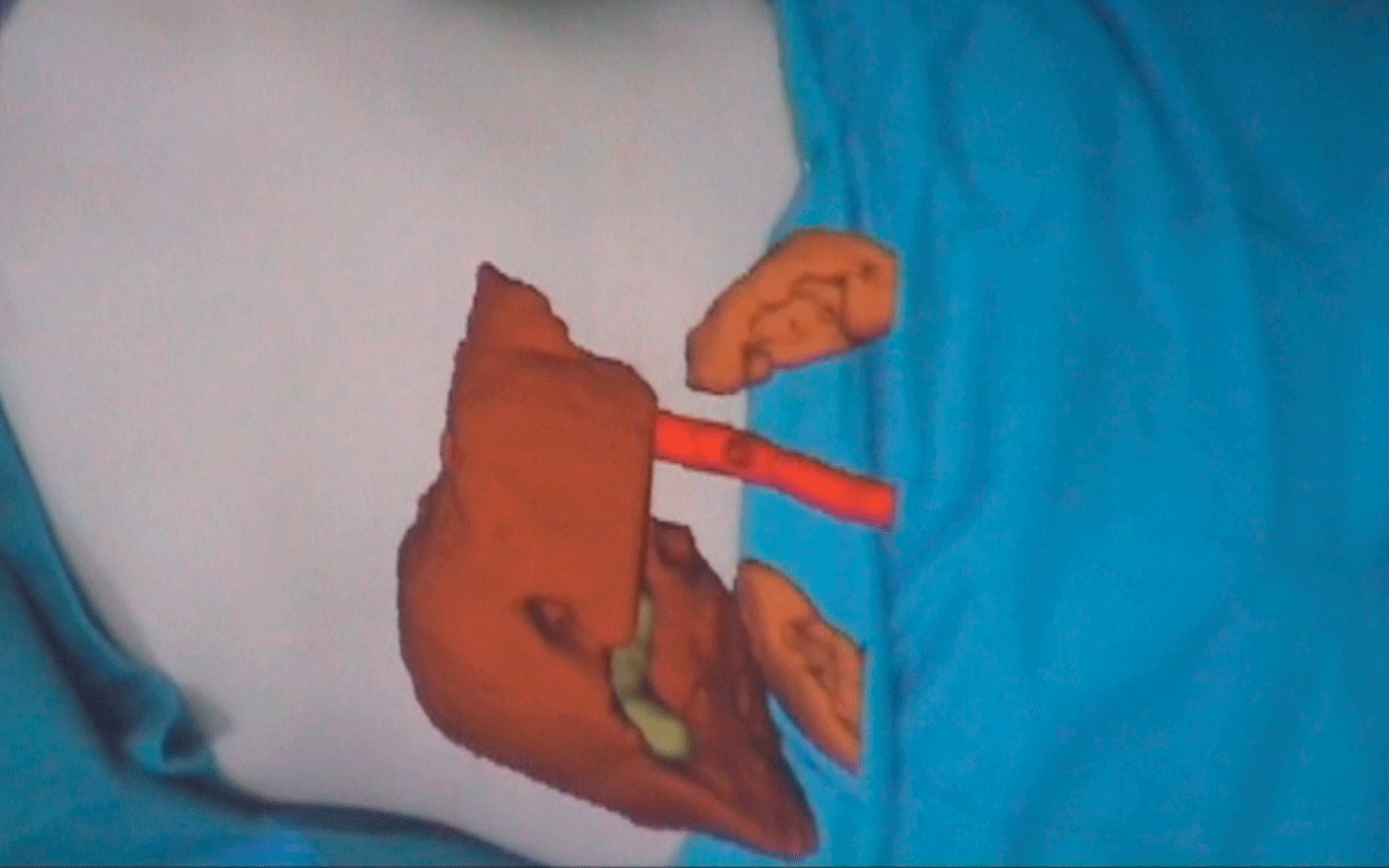
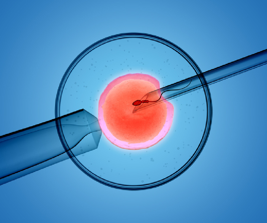
The project proposes the development of an Artificial Intelligence–based system capable of evaluating blastocysts in a completely non-invasive manner. To achieve this, multiple sources of information—traditionally analyzed separately—will be integrated. On the one hand, images and videos of embryo development obtained through time-lapse technologies will be used to study both morphology and temporal evolution. On the other hand, molecular data extracted from the culture medium, such as cell-free DNA and proteomic profiles, will provide key insights into the genetic and functional state of the embryo.
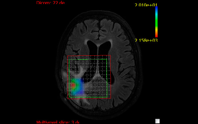
Based on this combination of data, advanced deep learning models will be developed to identify complex patterns and relationships that are not detectable using conventional methods. This approach will not only enable the determination of embryo ploidy without the need for biopsy, but also improve the prediction of implantation potential and treatment success, providing specialists with an objective and highly valuable decision-support tool.
One of the most innovative aspects of the project lies in the fusion of multimodal information within a single Artificial Intelligence model, an approach that has not yet been explored in this field. This strategy opens the door to a more comprehensive and accurate evaluation of embryos, reducing the uncertainty associated with current methods and enabling improved embryo selection.
The expected impact is twofold. From a clinical perspective, the project aims to increase success rates in assisted reproduction treatments, reduce risks associated with invasive techniques, and improve patient experience. From a technological standpoint, it will contribute to advancing the state of the art in Artificial Intelligence applied to biomedicine, generating new methodologies and capabilities that can be transferred to other domains.
NAIAEANIBL is being developed through a collaboration between IVI Valencia, an international leader in assisted reproduction providing the clinical environment and medical expertise for validation, and the Universitat Politècnica de València (UPV), through the CVBLab research group, which leads the development of the Artificial Intelligence models based on its expertise in computer vision and advanced biomedical data analysis.
Client

![]()

Partners